Reversible Cerebral Vasoconstriction Syndrome
Recognition, diagnostic pitfalls, delayed stroke, imaging and management in the emergency department
Introduction
Thunderclap headache represents a potential emergency that requires investigation. The immediate concern is aneurysmal subarachnoid haemorrhage. Second to this, is reversible cerebral vasoconstriction syndrome (RCVS).
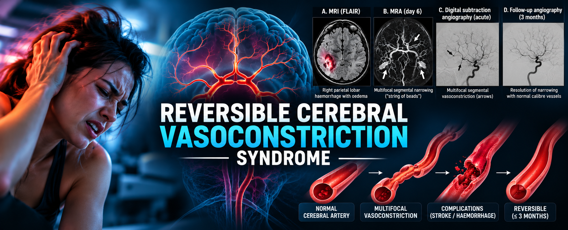
RCVS is characterised by sudden, severe headaches and reversible narrowing of intracranial arteries. It is a self limiting disease and most patients recover. However complications can include subarachnoid haemorrhage, intracerebral haemorrhage, cerebral infarction and posterior reversible encephalopathy syndrome (PRES).
What is RCVS?
RCVS is a syndrome involving transient dysregulation of cerebral vascular tone. The actual pathophysiology is still not well understood.
The characteristic findings are:
- Acute severe headache, described as thunderclap in 85% of RCVS cases.
- Recurrent thunderclap episodes over days or weeks.
- There is a characteristic pattern of repeated episodes, often averaging four thunderclap headaches over one to four weeks.
- Severe episodes commonly last one to three hours, although duration varies.
- A moderate headache may persist between episodes.
- There is often no neurology to find on clinical examination, unless there has a been a complication of infarction or haemorrhage.
- The original CT/CTA may be normal.
- Multifocal segmental narrowing of intracranial arteries is seen on angiography.
- The CSF is normal or near-normal in typical cases, although there may be a slight increase in both white blood cells and protein.
- Substantial or complete resolution of vascular abnormalities occurs within approximately 12 weeks. This makes the diagnosis.
RCVS can occur spontaneously or in association with triggers such as vasoactive medications, recreational substances or postpartum hormonal changes.
Triggers
It is important to specifically ask about triggers.
Common triggers include:
- Sexual intercourse, (the headache occurs just before, or at orgasm)
- Physical exertion
- Coughing or sneezing
- Defecation and straining
- Bathing or showering
- Emotional stress
There is an association between RCVS and vasoactive substances.
| Category | Examples |
|---|---|
| Recreational drugs | Cannabis, cocaine, amphetamines, MDMA |
| Antidepressants | SSRIs, SNRIs |
| Migraine medications | Sumatriptan, other triptans, ergot derivatives |
| Sympathomimetics | Pseudoephedrine, ephedrine |
| Other associations | Postpartum state, catecholamine-secreting tumours, certain immunosuppressive treatments |
These exposures may contribute to altered cerebrovascular tone.
Migraine and RCVS may share several symptoms, including nausea, vomiting, photophobia and phonophobia. However, the speed of onset is a crucial distinction. A key clinical issue is that triptans and ergot derivatives, frequently prescribed for migraine, can worsen cerebral vasoconstriction. They should be avoided when RCVS is suspected.
In more than 2/3 of cases RCVS occurs in the post partum period, usually within 1 week post delivery. It may follow a normal pregnancy, or one complicated by proteinuria or HELLP Syndrome. More than 1/3 of patients have been exposed to vasoconstrictoirs used in epidurals, or post partum haemorrhage.
Presentation
One of the most important features of RCVS is that symptoms, vascular abnormalities and neurological complications do not necessarily occur simultaneously.
Early presentation
Patients may initially have severe thunderclap headache with:
- Normal neurological examination ( normal neurological examination does not rule out the serious causes of a thunderclap headache)
- Normal non-contrast CT
- Normal MRI
- Normal early vascular imaging
In one study up to 55% had a normal initial CT or MRI, while 81% had brain lesions were identified during the course of the illness ie., they were delayed.
Evolution over time
This may be a clinical sequence of events:
| Time from onset | Typical manifestations |
|---|---|
| Days 0–3 | Thunderclap headache; imaging may be normal |
| First week | Recurrent headache, convexity SAH, intracerebral haemorrhage or PRES |
| Second week | Increasing vasoconstriction and delayed cerebral infarction |
| Weeks 2–3 | Vasoconstriction often reaches maximum severity |
| Up to 12 weeks | Substantial or complete resolution of vascular narrowing |
This timeline has important clinical implications:
- A patient may develop cerebral infarction after the initial headache has improved or resolved.
- Early clinical improvement therefore does not necessarily indicate that the cerebrovascular process has resolved.
Neurological complications
 Convexity subarachnoid haemorrhage
RCVS is associated with non-aneurysmal convexity SAH. Unlike aneurysmal SAH, which commonly involves basal cisterns, RCVS-related bleeding usually occurs within superficial cortical sulci and may be subtle on non-contrast CT(detected in 50% of cases). MRI, including FLAIR and susceptibility-sensitive sequences, may improve detection.
Intracerebral haemorrhage
Intraparenchymal haemorrhage is an important complication. These haemorrhages are usually lobar(more superficial) and single and are often associated with other types of stroke. They tend to occur early but may be delayed for several days.
Ischaemic stroke
Cerebral infarction typically occurs later (approximately 9 days) than haemorrhagic complications. This reinforces the need to recognise RCVS early and arrange appropriate surveillance.
Infarcts frequently involve watershed regions.
Posterior reversible encephalopathy syndrome (PRES)
RCVS may overlap with PRES. PRES is characterised by vasogenic cerebral oedema and may present with seizures, altered consciousness, visual symptoms and headache.
Although cerebral oedema often resolves, severe cases may develop infarction or haemorrhage.
The RCVS₂ score
The RCVS₂ score, a diagnostic tool developed to help distinguish RCVS from other intracranial arteriopathies. However , the presence of a thunderclap headachache alone requires that we consider the diagnosis.
The score incorporates five clinical and imaging features:
| Feature | Points |
|---|---|
| Thunderclap headache | +5 |
| Intracranial carotid artery involvement | −2 |
| Vasoactive trigger | +3 |
| Female sex | +1 |
| Subarachnoid haemorrhage | +1 |
The maximum score is 10.
- A score of 5 or greater had 90% sensitivity and 99% specificity for diagnosing RCVS.
- A score of 2 or less had 85% sensitivity and 100% specificity for excluding RCVS.
Clinical implication: The RCVS₂ score can support diagnostic reasoning, but it is not a replacement for appropriate imaging, investigation of dangerous alternative diagnoses or follow-up. I am not sure of the usefullness of the score or its applicability outside the population in which it was derived.
Diagnostic approach
Imaging
All patients presenting with thunderclap headache require urgent assessment for serious secondary causes.
The differential diagnosis includes:
- Aneurysmal SAH
- Intracerebral haemorrhage
- RCVS
- Cerebral venous sinus thrombosis
- Cervical artery dissection
- Pituitary apoplexy
- CNS infection
- Spontaneous intracranial hypotension
Non-contrast CT brain is generally an important first investigation, especially when the onset of headache has been less than 6 hours. Additional testing for SAH depends on the clinical presentation, CT timing and applicable diagnostic pathway.
 CTA and MRA
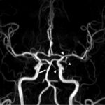
Vascular imaging may demonstrate multifocal segmental narrowing and dilatation, often described as a ‘string-of-beads’ appearance. However, early vascular imaging may be normal. Repeat imaging may be necessary.
MRI brain
MRI is valuable for identifying:
- Small cortical haemorrhages
- Acute cerebral infarction
- PRES
- Other intracranial abnormalities
Lumbar puncture
Up to 60% of patients may have some abnormalities in the CSF. These may include increased white blood cells and increased protein. If this is the case, repeat CSF sampling should be performed in a few weeks.
RCVS versus primary angiitis of the CNS
RCVS can resemble primary angiitis of the central nervous system (PACNS).
Distinguishing these conditions is important because treatment differs substantially.
| Feature | RCVS | PACNS |
|---|---|---|
| Onset | Sudden, often thunderclap | Usually insidious |
| Headache | Recurrent thunderclap | More often progressive |
| CSF | Usually normal or near normal | Often inflammatory |
| MRI | May initially be normal | Usually abnormal |
| Vascular abnormalities | Typically reversible | Often persistent or progressive |
| Steroid treatment | Generally avoided | May be required |
Repeat vascular imaging can be particularly helpful.
Improvement or resolution of arterial narrowing supports RCVS, whereas persistent or progressive abnormalities raise concern for alternative arteriopathies.
Management
Management remains largely supportive.
Remove potential triggers
Suspected vasoactive medications and recreational substances should be identified and withdrawn where appropriate. Triptans and ergot derivatives should be avoided.
Patients should temporarily avoid activities that provoke severe headaches during the acute phase.
Supportive treatment
Treatment includes analgesia, management of seizures, monitoring for neurological deterioration and appropriate management of haemorrhagic or ischaemic complications.
Blood pressure management requires care. Severe hypertension may require treatment, but hypotension may compromise cerebral perfusion in patients with significant vasoconstriction.
Nimodipine
Nimodipine is frequently used for symptomatic treatment and may reduce headache frequency or intensity.
Corticosteroids
Steroids have not demonstrated benefit and may be associated with clinical deterioration. This reinforces the importance of distinguishing RCVS from PACNS.
Prognosis and follow-up
Most patients experience resolution of headache and vascular abnormalities over several weeks. Patients who develop infarction or haemorrhage may have persistent neurological deficits and long-term prognosis depends primarily on the severity of cerebrovascular complications.
Follow-up imaging is important to confirm substantial or complete resolution of vasoconstriction, generally within approximately three months.
Patients also require counselling about potential triggers and recognition of recurrent neurological symptoms.
References
- Ducros A. Reversible cerebral vasoconstriction syndrome. Lancet Neurol. 2012;11(10):906–917.
- Son M, et al. Pearls & Oy-sters: A Journey Through Reversible Cerebral Vasoconstriction Syndrome: Sex, Drugs, and Headaches. Neurology. 2021;96(13):632–635. doi:10.1212/WNL.0000000000011194.
- Rocha EA, et al. RCVS₂ score and diagnostic approach for reversible cerebral vasoconstriction syndrome. Neurology. 2019;92–e647.