
The Aslanger pattern is a non-contiguous lead pattern identifying inferior STEMI
Guidelines require STE in two contiguous leads for localization( ie., anterior, lateral, inferior) and diagnosis of a STEMI.
However, what we need to remember is that the ECG is a two dimensional representaion of a three dimensional structure. When we look at the leads on the 3 dimensional heart, they are in fact adjacent in many circumstances.
Aslanger et al(1) looked at this and found the following 3 criteria, that identified a new ECG pattern:
- Any STE in lead III, but no other inferior leads (plus STE in aVR and V1)
- STD in any of leads V4 to V6 but not in V2, with a positive T wave
- ST in lead V1 higher than ST in V2
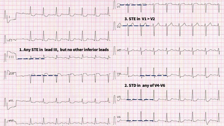
(1)
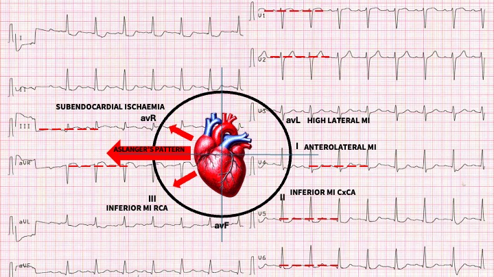
Modified from LITFL
We can consider this another OMI pattern. The significance is that it involves a significant blockage of the circumflex artery more than the right coronary artery. The fact that we don’t identify it as a STEMI, because of non-contiguous leads may result in significant loss of myocardium.
In Aslanger’s study the pattern was present in 13.3% of patients with an inferior MI. We do need to be cautious as the pattern was also present in 0.5% of those without an acute MI. This may be the result of previous injury.
The significance of this pattern lies in the fact that it includes a combination of lower MI and other critical ischaemia, resulting in a larger infact size and may result in higher mortality, when compared to patients with an inferior MI
Be aware of this pattern, however also be aware that in respect to the study:
-
This is a retrospective study with a potential for bias.
-
An isolated basal inferoseptal infarction or an acute inferior MI with previous infarctions may give a similar picture.
-
The pattern may be due to chronic change from a previous ischemic insult.
References
- Aslanger E et al. A new electrocardiographic pattern indicating inferior myocardial infarction. J of Electrocardiology. 61 (2020) 41–46