The Crashing PE Patient: What Works
Acute management of the crashing PE patient involves bridging therapies until clot resolution occurs. Although we have guidelines, individual approaches differ, which occurs when the literature is not entirely compelling.
For us to be able to make the right decisions for our patients and individualise treatment, we need to understand in more detail what happens physiologically in a massive PE, how we can assist that physiology, as well as to understand what each of the treatments provides.
I’ve just spoken on this topic in detail at EMCORE.
Why does the PE patient crash?
The patient with PE usually crashes because the right ventricle suddenly cannot pump blood through an acutely obstructed, high-resistance pulmonary circulation.
The problem starts in the pulmonary arterial circulation. A thrombus, usually originating in the deep veins, travels through via the IVC to the Right atrium(RA), then right ventricle(RV), then into the pulmonary artery and pulmonary arterial branches.
The physiological issue is twofold ie., how much functional pulmonary vascular bed has been lost (the degree of obstruction) and how much pulmonary vascular resistance (PVR) rises.
Pulmonary artery pressure begins to rise substantially when roughly 30–50% of the pulmonary arterial cross-sectional area is obstructed, however clot burden does not perfectly predict haemodynamic collapse. These other factors need to be taken into account.
So PVR rises through several mechanisms:
- Mechanical obstruction
- Inflammatory mediated vasoconstriction
- Release of vasoactive mediators, including thromboxane A₂ and serotonin, as well as, endothelial dysfunction and platelet activation contribute to pulmonary vasoconstriction and vascular dysfunction.
- Hypoxic pulmonary vasoconstriction
The Right Ventricle
The right ventricle is thin walled, perfused in both systole and diastole and ejects blood into a low pressure , low resistance circulation. It tolerates volume(preload) well, but does not tolerate afterload.
When pulmonary vascular resistance rises, the afterload the right ventrical works against, also rises. There is an increase in right ventricular systolic pressure, which leads to RV dilatation and an increase in RV wall tension.
Sympathetic stimulation will increase the heart rate and the contractility initially, however this cannot be maintained and the RV cannot generate progressively larger pressures, so it begins to dilate and then fail.
As the RV dilates, these things happen simultaneously:
- RV dilation stretches the tricuspid annulus leading to tricuspid regurgitation and a decrease in effective forward RV output
- The enlarged, high-pressure RV pushes the interventricular septum toward the LV. This impairs LV filling and as follows left ventricular output.
- The pulmonary obstruction results in decreased left ventricular filling and thus left ventricular stroke volume and cardiac output, resulting in hypotension.
Hypotension and the RV
This creates perhaps the downward spiral in crashing PE.
As the RV dilates and expands, there is increased wall tension and increased oxygen demand. The concurrent resulting hypotension also results in a decrease in right coronary artery perfusion.
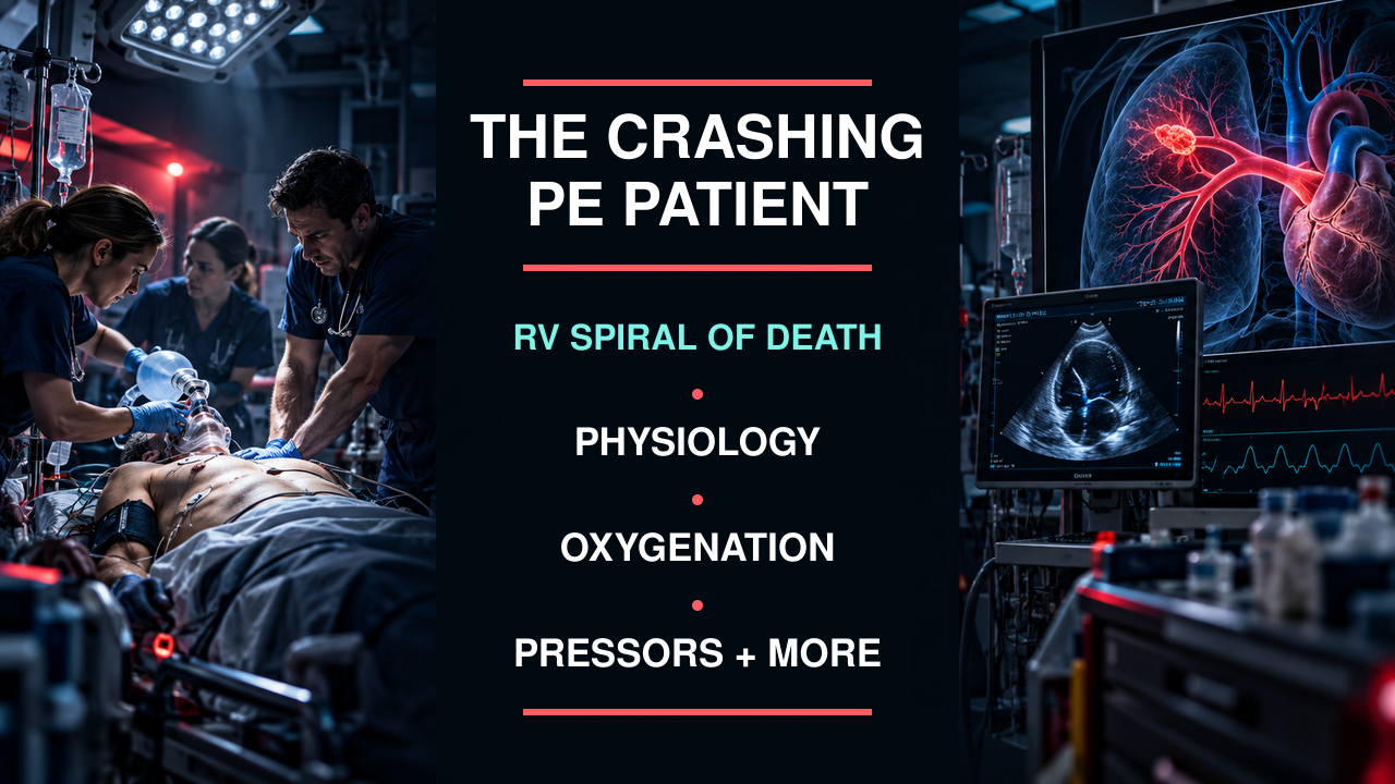
This causes RV ischaemia, decreasing RV contractility and RV output, which results in a drop in systemic BP and a decrease in coronary perfusion, which further increases the RV ischaemia. This is often called, the right ventricular spiral of death.

Hypoxaemia and Acidosis
PE creates a V/Q mismatch, so there are regions of lung that are ventilated but inadequately perfused. This contributes to hypoxaemia. Hypoxaemia results in increased PVR, increasing RV afterload which worsens output.
A fall in cardiac output results in a decrease in systemic perfusion resulting in tissue hypoxia and anaerobic metabolism, producing lactate, which results in a metabolic acidosis. Hypercapnoeia may add a respiratory acidosis to that.
Acidaemia can:
- increase PVR
- reduce myocardial contractility and responsiveness to catecholamines.
This is why a rapidly increasing lactate is a very important part of clinical assessment.
A word on Tachycardia
Intitially tachycardia is a compensatory response to maintain cardiac output. However excessive tachycardia can decrease diastolic filling and coronary perfusion time, whilst simultaneously increasing myocardial oxygen demand.
Therefore the compensatory sympathetic response can eventually contribute to RV ischaemia and failure.
What are we trying to achieve in these patients?
In the crashing PE patient, we must raise the blood pressure and oxygen saturation. However the purpose of what we do is to break the RV death spiral by:
- reducing RV afterload
- maintain RV coronary perfusion
- preserve appropriate preload
- avoid interventions that worsen RV function
- and remove the obstruction to the circulation.
How do we do this?
- REDUCE RV AFTERLOAD
This definitive treatment is to remove the clot which may involve one or more of:
- systemic thrombolysis
- catheter-directed therapy
- mechanical thrombectomy
- surgical embolectomy.
Everything else we do in the unwell PE patients, is largely buying time for this to occur.
- CORRECT FACTORS ADDING TO THE RV AFTERLOAD
Correct/avoid factors that increase PVR:
- Hypoxaemia
- Hypercapnia
- Acidaemia
- OPTIMISE RV PRELOAD
The RV needs enough filling to generate output, but not more than that as a dilated RV can lead to decreased cardiac output by:
↑ RV dilation → ↑ TR → ↑ septal shift → ↓ LV filling → ↓ cardiac output.
We need to take care with volume expansion. We need adequate preload, not maximum preload.
The current guideline supports volume expansion when there is clinical evidence of reduced preload; older ESC guidance similarly recommends cautious small-volume loading in selected patients rather than indiscriminate resuscitation.
- MAINTAIN SYSTEMIC PRESSURE AND RV CORONARY PERFUSION
The failing RV needs a sufficient systemic arterial pressure to maintain its coronary blood supply. We should:
- maintain MAP
- maintain RCA perfusion
- preserve RV contractility
- avoid xcessive airway pressures
- avoid intubation and positive-pressure ventilation, if possible.
The 2026 AHA PE guideline recommends vasopressors and/or inotropes in PE-associated cardiogenic shock to improve cardiac output and systemic perfusion.
Which vasopressor and/or inotrope is best. Keep reading.
TREATMENT
Hypoxaemia
The hypoxaemia in severe PE is primarily caused by V/Q mismatch, reduced pulmonary blood flow and low mixed venous oxygen saturation. Oxygen helps, but it doesn’t correct the fundamental obstruction.
Hypoxaemia itself can result in pulmonary vasoconstriction and increased PVR, which increases RV afterload and decreases RV output.
How to correct hypoxaemia
The new 2026 AHA/ACC PE guideline specifically states that for moderate–severe hypoxia, heated HFNC is beneficial compared with standard nasal oxygen, and recommends avoiding deep sedation and mechanical ventilation unless clinically necessary because both induction and positive-pressure ventilation can precipitate haemodynamic collapse.
HFNC gives a high FiO₂, washes out nasopharyngeal dead space, reduces work of breathing, and provides only relatively modest positive airway pressure. That makes it particularly attractive when the RV is impaired.
Move early to heated HFNC rather than repeatedly escalating conventional nasal oxygen. If HFNC is inadequate, a cautious trial of low-level NIV can be considered in selected patients, however care should be taken about PEEP/CPAP because increasing intrathoracic pressure can reduce venous return and RV preload while increasing RV afterload.
Beware Intubation and even try and avoid it, unless you have a patient with refractory profound hypoxaemia, exhaustion/inability to maintain ventilation, inability to protect the airway, severe altered consciousness, or arrest/peri-arrest circumstances
Low saturation + severe tachypnoea does not automatically equal intubation.
The negative effects of intubation?
The patient may be maintaining circulation through:
high sympathetic tone + spontaneous negative-pressure breathing + venous return.
Intubation affects the very things that are maintain circulation leading to peri/post intubation cardiac arrest rates of 8-12% (4 times the normal emergency intubation rate):
- Sedation/Induction removes sympathetic tone, decreasing SVR
- positive-pressure/PEEP ventilation increases intrathoracic pressure, decreases venous return and decreased RV preload, resulting in decreased cardiac output, with subsequent hypotension.
When intubation is unavoidable, treat this as a high-risk haemodynamic procedure, not an ordinary RSI.
- Have haemodynamic rescue established or immediately available before induction—particularly vasopressor support—and have the team prepared for rapid deterioration. I have vasopressor support running aiming for a systolic blood pressure of 120-140 mmHg.
- Once ventilated, avoid excessive airway pressures. Older ESC guidance recommends approximately 6 mL/kg lean body weight, plateau pressure <30 cm H₂O, and beware of PEEP, because positive intrathoracic pressure can reduce venous return and worsen low cardiac output.
- Avoid allowing major hypercapnia/acidaemia if possible, because both can increase PVR and further load the RV.
Refractory hypoxaemia is certainly telling us that pulmonary blood flow is catastrophically compromised.
TREATING HYPOTENSION
What would the ideal drug look like, for the crashing PE patient?
The ideal drug/combination of drugs should:
- raise systemic pressure,
- improve RV coronary perfusion
- improve RV contractility,
- Not increasing—and preferably decrease—PVR.
No single drug does all of this perfectly. The 2026 PE guideline recommends vasopressors/inotropes for PE-associated cardiogenic shock and considers noradrenaline the usual first vasopressor. Recommendations are made for second line agents which may include Vasopressin or an Inodilator.
Here are some of the drugs used
| Class | Drug | α1 | β1 | β2 | Other | SVR | PVR | Contractility | Main PE role |
| INOPRESSOR | Noradrenaline | +++ | ++ | +/0 | — | ↑↑ | ↔ / ↑* | ↑ | First-line pressor |
| INOPRESSOR | Adrenaline | ++–+++ | +++ | ++ | — | ↑↑† | ↔ / ↑ | ↑↑ | Profound/refractory shock, peri-arrest |
| VASOPRESSOR | Vasopressin | 0 | 0 | 0 | V1 +++ | ↑↑ | ↔ / ↓‡ | ↔ | Add when noradrenaline requirement escalating |
| INODILATOR | Dobutamine | 0/+ | +++ | ++ | — | ↓ / ↔ | ↓ | ↑↑ | Low CO despite adequate BP |
| INODILATOR | Milrinone | 0 | 0 | 0 | PDE-3 inhibitor | ↓↓ | ↓ | ↑↑ | Selected RV failure when BP can tolerate vasodilation |
The guidelines recommend starting with Noradrenaline in the crashing PE patient. However there are differing opinions of the right approach.
Remember that the use of medications is a bridging therapy and in a situation, where lysis is your only option, that may have already been commenced, shilst the blood pressure is being stabilised.
Inopressors and beta and alpha effects?
α1: Mainly causes vasoconstriction
β1: Affects heart rate and contractility
β2: Affects vasodilatation including that of the pulmonary vasculature
Adrenaline
| Adrenaline dose* | Predominant effects | β1 | β2 | α1 | Typical haemodynamic result |
| ~0.01–0.05 μg/kg/min | Predominantly β | ↑↑ | ↑↑ | ↑ | ↑ HR, ↑ contractility, β2 vasodilation |
| ~0.05–0.1 μg/kg/min | Mixed α + β | ↑↑↑ | ↑↑ | ↑↑ | ↑ CO, increasing SVR |
| ~0.1–0.2 μg/kg/min | Strong mixed effect | ↑↑↑ | ↑↑ | ↑↑↑ | ↑ BP, ↑ CO, marked inotropy |
| >~0.2 μg/kg/min | Increasingly α-dominant | ↑↑↑ | ↑/↑↑ | ↑↑↑↑ | marked vasoconstriction + inotropy |
Low dose Adrenaline:
Adrenaline has predominantly beta effects at lower doses and becomes more alpha as the dose increases. At doses up to 0.05 mcg/kg/min it is predominantly beta ie., for a 70 kg patient 3.5 mcg/ min. When this dose is increased to above 0.1 mcg/kg/min ie., 7 mcg/min, the alpha effects are becoming dominant.
β1 + β2 > α1
β1: increases cardiac contractility and heart rate resulting in increased cardiac output
β2: causes systemic vasodilation
Consequently, at very low doses cardiac output increases but SVR may fall or change relatively little.
This is why low-dose adrenaline can behave more like an inotrope/inodilator than a pure pressor.
Intermediate dose adrenaline
α1 activity becomes increasingly important.
Given the β1 activity leading to increased contractility and α1 leading to increased vasoconstriction adrenaline results in increased CO, SVR and MAP.
High-dose adrenaline
At doses above 0.2 mcg/kg/min ie., >14 mcg/min, the effects are predominantly alpha, however they do include beta effects. There is variability from patient to patient.
α1 vasoconstriction becomes increasingly dominant and β1 stimulation still persists.
The increased SVR and MAP, as well as heart rate and contractility results in increased myocardial oxygen consumption which can have deleterious effects on a RV struggling with afterload.
Noradrenaline
Noradrenaline is different. It is predominantly an alpha drug even at low doses, with minimal beta2 effects throughout it’s whole dose range.
Noradrenaline has:
α1 +++ / β1 ++ / very little β2
So unlike adrenaline, there isn’t a clinically useful transition from a “β drug” into an “α drug.” We are predominantly increasing the magnitude of α-mediated vasoconstriction rather than changing from β-dominant to α-dominant physiology.
It is already substantially α-mediated at low doses.
| Noradrenaline dose* | α1 | β1 | β2 | Dominant effect |
| ~0.01–0.05 μg/kg/min | ↑↑ | ↑ | minimal | Vasoconstriction + some inotropy |
| ~0.05–0.1 μg/kg/min | ↑↑↑ | ↑↑ | minimal | ↑ SVR/MAP + β1 support |
| ~0.1–0.2 μg/kg/min | ↑↑↑ | ↑↑ | minimal | Strong vasopressor/inopressor |
| >0.2 μg/kg/min | ↑↑↑↑ | ↑↑ | minimal | Increasing vasoconstriction |
What do we want to achieve in the crashing PE patient?
We want to increase and increase SVR and aortic pressure which increases right coronary perfusion, thus support RV myocardium. At the same time we don’t want increased PVR but decrease it, as this leads to increased RV afterload and worsening RV failure.
Our objective in the crashing PE isn’t simply to produce vasoconstriction. We want to increase systemic pressure enough to perfuse the RV, while avoiding excessive pulmonary vasoconstriction that further increases RV afterload.
So an approach is to start noradrenaline → add vasopressin and/or dobutamine according to whether the remaining problem is pressure or flow.
How do the drugs affect this?
Noradrenaline
At modest doses Noradrenaline can increase SVR substantially without proportionately increasing PVR.
The 2026 PE guideline makes the interesting observation that noradrenaline at ≤15 μg/min has little effect on PVR, producing a favourable increase in the SVR:PVR ratio. At higher doses, PVR may begin to increase.
Increasing the dose, gives a progressively greater α1 vasoconstriction. This results in increased SVR and MAP, which leads to increased RV coronary perfusion
However eventually this may result in increased PVR, potentially increasing RV afterload
This is the physiological rationale for adding another agent rather than escalating noradrenaline.
Adrenaline
At lower doses β1/β2 effects are prominent resulting in increased RV contractility, CO with some β2 vasodilation
At intermediate doses alpha effects increase, so we have β1 + α1, which result in increased contractility, SVR and MAP.
At high doses there is increasing α1 dominance, with significant increases in SVR and potentially PVR. There is increased myocardial oxygen consumption and tachycardia and an increased arrhythmia risk.
Summary of Adrenaline and Noradrenaline
| NORADRENALINE | ADRENALINE | |
| Low dose | α1 > β1 | β1/β2 > α1 |
| Moderate dose | α1 ↑↑ + β1 | β1 + α1 ↑↑ |
| High dose | α1 increasingly dominant | α1 increasingly dominant + strong β1 |
| β2 | Minimal | Important at low dose |
| SVR | ↑ from low dose | ↔/↓ → ↑ → ↑↑ |
| Contractility | ↑ | ↑↑ |
| HR | ↔/slight ↑ | ↑↑ |
| PVR | ↔ initially; may ↑ at high dose | Variable; concern with high doses |
| PE role | First-line pressor | Profound/refractory shock or peri-arrest |
The 2026 AHA/ACC PE guideline supports exactly this distinction: above ~15 μg/min, noradrenaline may begin to increase PVR, so if hypotension persists, add a second vasopressor; if cardiac output remains low despite vasopressor support, add an inotrope.
| At NE >15 μg/min | Add VASOPRESSIN | Add DOBUTAMINE |
| Main problem | Pressure | Flow / RV pump failure |
| MAP | Still low | Adequate or near adequate |
| SVR | Inadequate | Often adequate/high |
| Cardiac output | May be adequate for circumstances | Low |
| Lactate | May be ↑ | Often ↑ / continuing to rise |
| Extremities | Variable | Often cold/poor perfusion |
| Urine output | ↓ | ↓ |
| Echo | RV dysfunction | Severe RV dysfunction + poor forward flow |
| LV | May be underfilled | Markedly underfilled |
| Effect wanted | ↑ SVR/MAP | ↑ RV contractility/CO |
| PVR effect | relatively neutral at low dose | ↓ |
| Major risk | Excess vasoconstriction | Hypotension |
VASOPRESSIN
Vasopressin has no α or β activity. It activates V1 receptors which produce vascular smooth-muscle constriction through a completely different pathway from catecholamines.
It results in increased SVR and a decrease in PVR at low doses, but no effect on heart rate or contractility.
This makes vasopressin attractive when you need more systemic pressure but don’t want simply to keep increasing adrenergic stimulation. Contemporary RV-failure literature reports increased SVR with relatively favourable effects on PVR.
INODILATORS: pump harder against less resistance
These become particularly interesting in PE as they can increase contractility and decrease pulmonary vascular resistance. However they also decrease SVR and can cause hypotension and decreased right coronary perfusion.
Dobutamine
β1 +++ : β2 ++ : α1 0/+
There is predominant β1 (+++) activity and thus they:
- Increase RV contractility
- Increase stroke volume
- Increase cardiac output
β2 ++ effects include:
- systemic vasodilation
- pulmonary vasodilation
There is weak α activity, which doesn’t compensate sufficiently for the vasodilation.
Therefore it has the following effects:
- CO ↑↑
- PVR ↓
- SVR ↓/↔
- BP ↓/↔
- HR ↑
Low-dose dobutamine increases CO while reducing PVR and filling pressures;
At 5–15 μg/kg/min it produces a larger increase in CO but reduces SVR.
Milrinone
Milrinone is a PDE-3 inhibitor so has no α or β receptor activity. It increases intracellular cAMP. In the myocardium it results in increased Ca²⁺ availability, resulting in increased contractility.
In vascular smooth muscle, it results in vasodilation
It has the following effects:
- Contractility ↑↑
- CO ↑↑
- PVR ↓
- SVR ↓↓
The acute-RV-failure literature describes milrinone as increasing CO and reducing PVR, but producing an even greater reduction in SVR.
That makes it physiologically attractive for the RV but potentially dangerous in the hypotensive crashing PE patient.
| INOPRESSORS | VASOPRESSOR | INODILATORS | |
| Noradrenaline | Vasopressin | Dobutamine | |
| Adrenaline | Milrinone | ||
| Primary goal | PRESSURE + PUMP | PRESSURE | PUMP + ↓ AFTERLOAD |
| α1 | NA +++ / Adr ++–+++ | 0 | Dob 0/+ / Mil 0 |
| β1 | NA ++ / Adr +++ | 0 | Dob +++ / Mil 0 |
| β2 | NA +/0 / Adr ++ | 0 | Dob ++ / Mil 0 |
| Other | — | V1 +++ | Milrinone: PDE-3 |
| SVR | ↑↑ | ↑↑ | ↓ |
| PVR | ↔/↑ | ↔/↓ | ↓ |
| CO | ↑ | ↔ | ↑↑ |
An Approach
In the patient being treated with noradrenaline for hypotension:
- If the MAP is still low ie., 50–60 mmHg, despite noradrenaline give vasopressin
- If the MAP is fine ie., 70 mm Hg, but the patient is still shocked give dobutamine
Case Examples
Case 1
Patient has been commenced on Noradrenaline and is at 18 μg/min
The BP 78/45, MAP 56
On bedside echo the RV dilated, patient shocked.
There is INSUFFICIENT SYSTEMIC PRESSURE We need systemic arterial pressure to maintain RV coronary perfusion.
VASOPRESSIN can be added to increase SVR and MAP and thus right-coronary perfusion and RV without simply pushing noradrenaline higher and potentially increasing PVR. This is specifically the approach suggested in the 2026 guideline.
Case 2
The patient is on 18 μg/min of Nadr and has a BP 95/60 with a MAP of 72
However, the following findings, indicating the patient is still shocked:
lactate 6 → 8 mmol/L
cold peripheries
oliguria
poor mentation
echo: huge poorly contracting RV
tiny LV / poor LV filling
The problem here is not a need for more vasoconstriction, but for more forward flow:
Add DOBUTAMINE which will increase RV contractility and decrease PVR, resulting in increased RV output and pulmonary blood flow, which will increase LV preload, LV stroke volume and cardiac output.
The guideline specifically states that dobutamine may be added to noradrenaline in hypotensive PE when cardiac output remains low, although it can lower SVR.
How do I determine “low flow” at the bedside?
Look for:
Clinical perfusion: altered mentation, cold/mottled peripheries, prolonged capillary refill, oliguria.
Metabolic: rising/persistently elevated lactate and metabolic acidosis.
Echo: severely dilated RV, poor RV systolic function, septal flattening/bowing, severe TR, underfilled LV and poor LV stroke volume.
If available, haemodynamic measures can strengthen the diagnosis: cardiac index ≤2.2 L/min/m² is one of the guideline’s markers of hypoperfusion/normotensive shock.
What if BOTH pressure and flow are bad?
With NAdr >15 μg/min and the MAP is 55 mmHg + lactate rising + severe RV failure + low output.
In this case we may need to add both VASOPRESSIN and DOBUTAMINE
Inhaled Pulmonary Vascular Dilators
In the crashing PE patient, inhaled pulmonary vasodilators are particularly attractive because they can reduce PVR and RV afterload without producing the systemic hypotension associated with IV pulmonary vasodilators. The new 2026 AHA/ACC PE guideline says they may be considered in PE Categories C2–E to reduce RV afterload.
| Agent | Mechanism | Typical acute dose | PVR | SVR/BP | Oxygenation | Practical ED role |
| Inhaled nitric oxide (iNO) | NO → ↑ cGMP → pulmonary vasodilation | 5–40 ppm | ↓↓ | Usually ↔ | ↑ | Best-established option |
| Inhaled epoprostenol | Prostacyclin/PGI₂ → ↑ cAMP | 10–50 ng/kg/min | ↓↓ | Usually ↔/↓ | ↑ | Excellent alternative to iNO |
| Inhaled iloprost | Prostacyclin analogue → ↑ cAMP | Often 2.5–5 μg nebulised | ↓ | Usually ↔ | ↑ | Longer acting; less convenient for crashing patient |
| Inhaled treprostinil | Prostacyclin analogue | Device/formulation dependent | ↓ | Usually ↔ | ↑ | More chronic PH than ED rescue |
| Nebulised nitroglycerin | NO donor → ↑ cGMP | Protocols vary; evidence limited | ↓ | Potential ↓ | ? | Rescue option when iNO/epoprostenol unavailable |
The availability of these may be the limiting step.
Inhaled Nitric Oxide(NO)(5-40 ppm) is used in ICU environments. It diffuses into the pulmonary vascular smooth muscle resulting in increased cGMP which produces smooth-muscle relaxation. This decreases PVR and decreases RV afterload
Its systemic vascular effect is minimal as it is rapidly inactivated by haemoglobin, on entering the bloodstream.
Nebulised GTN
Nebulised GTN is converted to pulmonary NO, which results in increased cGMP, which decreases PVR and thus RV afterload, increasing RV output.
This is an inhaled medication, not to be given IV. Dosage proposed is 5 mg nebulised over about 15–30 minutes, repeat as required for massive PE/RV failure. If the concentrated preparation isn’t available, nebulising 3–5 mL of the available IV GTN solution, although this delivers a smaller dose with dilute preparations. Reported onset is within minutes and effect lasts roughly 20–30 minutes.
The drug is delivered to ventilated lung units and may also improve V/Q matching.
References
- Creager MA, Barnes GD, Giri J, et al. 2026 AHA/ACC/ACCP/ACEP/CHEST/SCAI/SHM/SIR/SVM/SVN Guideline for the Evaluation and Management of Acute Pulmonary Embolism in Adults. J Am Coll Cardiol.2026;87(13):1626–1710.
This is a main reference. It supports contemporary risk classification, haemodynamic support, noradrenaline as preferred vasopressor, the >15 μg/min issue, addition of a second vasopressor, dobutamine for persistent low output, cautious fluids, inhaled pulmonary vasodilators and advanced PE therapies.
2026 Acute PE Guideline – JACC - Konstantinides SV, Meyer G, Becattini C, et al. 2019 ESC Guidelines for the diagnosis and management of acute pulmonary embolism. Eur Heart J. 2020;41:543–603.
Excellent reference for high-risk PE, RV failure, cautious fluid administration, noradrenaline, dobutamine, thrombolysis, surgical embolectomy, catheter treatment and ECMO. The ESC haemodynamic approach strongly complements the newer 2026 guideline. - Arrigo M, et al. Acute right ventricular failure: pathophysiology, aetiology, assessment, and management. Eur Heart J. 2025;46:2520–2544.
This is one of the best contemporary references for the RV death spiral - Dudzinski DM, Giri J, Rosenfield K. Interventional treatment of pulmonary embolism. Circ Cardiovasc Interv.2017;10:e004345.
Useful background for the transition from haemodynamic support to definitive reperfusion and catheter-based intervention. - Harjola VP, Mebazaa A, Čelutkienė J, et al. Contemporary management of acute right ventricular failure: a statement from the Heart Failure Association and Working Group on Pulmonary Circulation and Right Ventricular Function of the ESC. Eur J Heart Fail. 2016;18:226–241.
Excellent physiology reference for acute RV failure, ventricular interdependence, preload, afterload, vasopressors and inotropes. - Sifuentes AA., et al Mechanical Circulatory Support and Critical Care Management of High-Risk Acute Pulmonary Embolism. Interv Cardiol Clin. 2023 Apr 27;12(3):323–338. Contemporary review of high-risk PE physiology and critical care. It describes increased RV wall tension and oxygen demand, impaired RV coronary perfusion, RV dilatation, septal shift and subsequent reduction in LV preload and cardiac output.
- Piazza G, Goldhaber SZ. The acutely decompensated right ventricle: pathways for diagnosis and management. Chest. 2005;128:1836–1852.
- Haddad F, Doyle R, Murphy DJ, Hunt SA. Right ventricular function in cardiovascular disease, Part II: pathophysiology, clinical importance, and management of right ventricular failure. Circulation. 2008;117:1717–1731.
- Jardin F, Genevray B, Brun-Ney D, et al. Dobutamine: a hemodynamic evaluation in pulmonary embolism shock. Crit Care Med. 1985;13:1009–1012.
Small but important physiological study showing improved cardiac index with dobutamine in PE; it is specifically cited by the 2026 guideline. - De Backer D, Biston P, Devriendt J, et al. Comparison of dopamine and norepinephrine in the treatment of shock. N Engl J Med. 2010;362:779–789.
Supports the preference for noradrenaline over dopamine in shock and is incorporated into the 2026 PE guideline evidence base. - Levy B, Clere-Jehl R, Legras A, et al. Epinephrine versus norepinephrine for cardiogenic shock after acute myocardial infarction. J Am Coll Cardiol. 2018;72:173–182.
Useful when discussing why adrenaline is generally not the routine first choice when noradrenaline can achieve the required systemic pressure. - Mathew R, Di Santo P, Jung RG, et al. Milrinone as compared with dobutamine in the treatment of cardiogenic shock. N Engl J Med. 2021;385:516–525.
Not PE-specific, but useful evidence for comparing the two inodilators. It is also included in the 2026 PE guideline’s haemodynamic pharmacotherapy evidence. - Mercat A, Diehl JL, Meyer G, et al. Hemodynamic effects of fluid loading in acute massive pulmonary embolism. Crit Care Med. 1999;27:540–544.
Important primary reference behind the recommendation for small/cautious fluid challenges rather than aggressive volume loading. It is specifically included in the new guideline evidence base. - Lee ES, Baltsen CD, Stubblefield WB, et al. Intubation and Mechanical Ventilation in Patients with Acute Pulmonary Embolism: A Scoping Review. J Intensive Care Med. 2026. doi:10.1177/08850666241285862.
This directly addresses the danger of intubation in acute PE. Across the limited available studies, peri-intubation haemodynamic collapse occurred in 19–28%. - Advanced Cardiopulmonary Support for Pulmonary Embolism. Tech Vasc Interv Radiol. 2017.
Useful review emphasizing careful preload/vasoactive management, extreme caution around intubation and positive-pressure ventilation, and VA-ECMO as a bridge to reperfusion in selected patients. - Management of high-risk pulmonary embolism in the emergency department: A narrative review.
Useful ED-focused reference for oxygenation. It emphasizes improving oxygenation while avoiding unnecessary increases in intrathoracic pressure, using HFNC where appropriate, and avoiding NIV/intubation where possible in haemodynamically vulnerable patients. - Kline JA, Puskarich MA, Jones AE, et al. Inhaled nitric oxide to treat intermediate-risk pulmonary embolism: a multicenter randomized controlled trial. Nitric Oxide. 2019;84:60–68.
The iNOPE trial is the principal PE-specific randomized evidence for inhaled nitric oxide. It tested 50 ppm iNO in normotensive PE patients with RV dysfunction. - Kline JA, Hall CL, Jones AE, et al. Randomized Trial of Inhaled Nitric Oxide to Treat Acute Pulmonary Embolism: The iNOPE trial. Am Heart J. 2017;186:100–110.
Provides the physiological rationale and trial methodology; particularly useful for discussing pulmonary vasoconstriction as a contributor to increased PVR in PE.