Case Presentation:
During a recent shift as a VMO in an Eastern State Regional ED I was faced with a rarely encountered, clinically important and emerging clinical challenge.
Many of you would have noted the increase in media attention to the problem of tuberculosis, and particularly multi-drug resistant TB (MDR TB). 4 recent cases of MDR TB have been diagnosed in north Queensland. All the cases were imported from Papua New Guinea, particularly the Western Province of PNG via Daru island. The clinical significance of MDR TB is that the duration and cost of treatment are far greater than that of “traditional TB”, up to 24 months and $1 million by some estimates, though the financial aspect may involve some extrapolation.
The case that I encountered was of a lady who had moved to Australia some 7 years earlier from Manila, she denied any previous TB diagnosis and, one presumes, had undergone immigration medicals including chest x-ray. Her housemate had returned from a visit to the Philippines approximately 8 weeks before presentation and had been staying with a relative with TB. The patient had suffered from 4-6 weeks fevers, cough and weight loss of 4 kg (approximately 10% body weight). She had presented to her GP who had investigated her symptoms and included a Ziehl-Neesen (ZN) stain for acid fast bacilli (AFB). The initial stain was negative, but subsequent cultur was positive for AFB. She was referred to hospital via the ED.
We are not here to question the wisdom of referral via ED of a notifiable infectious disease, but merely to discuss how we manage these patients in ED.
The patient was flagged at triage by an astute triage nurse and urgent consultant review sought. I asked for the patient to be placed in the negative pressure room in Ed and universal precautions with PPE instituted for all those in contact with the patient. She was clinically well, although somewhat cachectic, and so could be managed appropriately in the isolation room. Confirmation of the microbiological diagnosis was sought from the external pathology lab and confirmation that the disease control unit had been notified for contact tracing was received.
The examination of the patient was essentially unremarkable. She was wasted, afebrile and cardiovascularly stable. Her respiratory system was unremarkable and she did not exhibit any lymphadenopathy.
Baseline haematology and biochemistry were normal and further sputum samples for ZN staining for AFB and extended culture and drug sensitivity testing were taken. Consent for HIV testing was obtained and the HIV test sent.
The patient underwent radiological investigation, she was transported by staff with PPE and wore an N95 mask at all times.
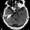
Chest X-Ray appearances were similar to those seen below:

A Gohn focus was noted on right with evidence of reactivated disease on the left. There is calcification of the hilar lymph nodes.
She was admitted under the general medical team team and started on a 4-drug regimen as advocated by World Health Organisation, awaiting infectious diseases input. The regimen is:
- 2HRZE/4HR
- 2 months of combination therapy isoniazid, rifampicin, pyrazinamide and ethambutol. Daily dosing preferable, but 3 times a week Direct Observed Therapy, Short course is acceptable (TB DOTS), as long as pt is not HIV +ve
- 4 months isoniazid and rifampicin. Daily dosing is preferable but TB DOTS 3 times a week is acceptable (1)
Discussion:
Tuberculosis is a bacterial infection, classically of the lungs, caused by the bacteria Mycobacterium tuberculosis. The illness is primarily spread through expectoration of respiratory droplets from a sick individual, and subsequent inhalation of said particles by exposed people. The organism then grows in the lungs in two main stages, depending on the immune status of the infected individual: latent infection and active disease. Latent TB occurs when the tuberculin skin test (TST) is positive, but the chest radiograph is normal and /or the patient is asymptomatic. Active disease occurs when there are clinical signs and symptoms of infection (pulmonary or extrapulmonary), and/or the CXR is abnormal Typical findings on a CXR of a patient with TB include hilar lymphadenopathy or Gohn complexes generally in the upper apices of the lungs.
TB is uncommon in Australia with annual incidences of 886 and 1,062 bacteriologically-confirmed cases of tuberculosis (TB) in 2008 and 2009, representing an annual rate of 4.1 and 4.9 cases per 100,000 population respectively.(2)
The commonest presentation is pulmonary TB. The patient is infected through droplet spread from an infected source. The initial infection rarely causes primary disease, the immune response ‘walls off’ the infection in the lungs through granuloma formation, the “Gohn complex”. Primary disease occurs in the immune-compromised and in infants. The commonest disease progression is that of quiescent disease. This disease may become reactivated due to acquired immune compromise or through re-infection.
The World Health Organisation (WHO) has a 4-symptom screen to assess for symptoms of TB:
- Fever (reported over last week or at time of consult)
- Night sweats (enough to require a change of shirt)
- Weight loss
- Current, ongoing cough lasting more than 2 weeks
2,3 or 4 positive symptoms in a high risk individual are sensitive, but not specific for pulmonary TB.
High risk historical factors include; previous treatment or diagnosis for TB, TB contacts, travel to a high prevalence region, HIV infection, institutional living and homelessness
The examination is often normal, but may demonstrate; lymphadenopathy, cachexia, nonspecific chest abnormalities. A low pitched wheeze over the granuloma is often cited, but in the 20+ cases of TB that I have seen in the last 2 years I have never detected that sign.
The laboratory diagnosis of TB is preferred prior to treatment initiation. (1) The gold standard is sputum culture. Direct ZN staining and traditional microscopy has sensitivity of about 20% whereas fluorescence microscopy is quoted as 30%+. A nucleic acid amplification test (NAAT), real time PCR, that identifies sections of TB DNA and takes 2 hours has been rolled out by the Bill and Melinda Gates foundation through southern Africa. This technology is known as the GeneXpert and is likely to change the face of TB diagnosis. The QuantiFERON Gold is an interferon gamma release assay that looks for markers of cellular response to TB infection, its clinical significance is that it can differentiate between latent infection and immunity through vaccination, unlike the Mantoux test.
The treatment of TB is reliant on the compliance of the patient for the duration of chemotherapy. The initial 2 month regimen of 4 drugs is recommended to be hospital based as inpatient in isolation as the patient remains contagious during this time. The subsequent dual therapy is outpatient based with direct observed therapy recommended if compliance is in any way in doubt. Poor compliance leads to the development of MDR TB or even XDR TB (extreme drug resistant TB).
15 cases of total drug resistant TB were report at the end of 2011 in India (3) (previous cases in Iran were unconfirmed) and may represent a totally new and terrifying disease challenge. We will be back in the same situation as we were in the late 19th and early 20th centuries when TB accounted for 20% adult deaths and there was nothing medicine could do about it.
Scary thought!
By Will Davies
- Treatment of Tuberculosis Guideline. WHO 2010. World Health Organisation, Geneva.
- Richard Lumb, Ivan Bastian, Robyn Carter, Peter Jelfs, Terillee Keehner, Aina Sievers. Tuberculosis in Australia: bacteriologically confirmed cases and drug resistance, 2008 and 2009. Communicable Diseases Intelligence Volume 35 No 2 – June 2011. http://www.health.gov.au/internet/main/publishing.nsf/content/cdi3502-1 Accessed 29/10/12
- Zarir F. Udwadia, Rohit A. Amale, Kanchan K. Ajbani, and Camilla Rodrigues. Totally Drug-Resistant Tuberculosis in India Clin Infect Dis. (2012) 54(4): 579-581