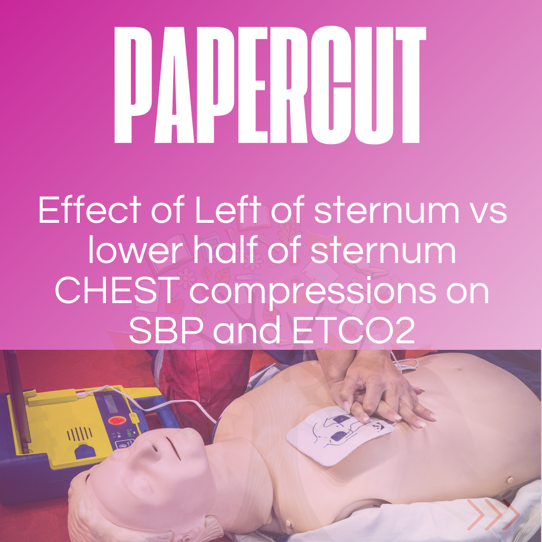
High Quality CPR, requires high quality chest compressions, that provide coronary artery and cerebral perfusion. The International Liaison Committee on Resuscitation (ILCOR) recommends chest compressions be performed in the lower half of the sternum. This is usually described as the internipple line. This however may not always be the best location as the position of the heart varies in individuals.
Shin (1) found that the left ventricle was at this level in only 20% of patients. Using these landmarks, it resulted in compression of the cardiac base (in up to 80% of cases), which included:
Transoesophageal ECHO used on intubated patients during cardiac arrest resuscitation (2), found that in 79% of cases, the area of maximal compression was within 2cm of the aortic valve. Compression of the left ventricular outflow tract resulted in narrowing of the aortic root and in some cases obliterated the aorta during compression.
The Study
Rolston D.M et al. Left of Sternum compressions are associated with higher systolic blood pressure than lower half of sternum compressions in cardiac arrest. Resuscitation 2024 ( Ahead of Print) doi: https://doi.org/10.1016/j.resuscitation.2024.110466
The Verdict Summary
This is a retrospective pilot study at a single centre.
The primary outcome was systolic blood pressure(SBP) and the secondary outcome, end-tidal CO2 (ETCO2). It doesn’t tell us anything about survival to hospital discharge, although 66.7% of patients who had their compressions changed to left of sternum achieved ROSC. Systolic rather than diastolic blood pressures were measured and interestingly, even though the SBP was higher with left of sternum compressions, there was no difference in ETCO2.
I’m not sure that this is meant to be a practice changing study. What it does, is highlight the need for tailored resuscitation as well as some of the limitiations of mechanical chest compressions.
The most important quote for me from this study is; “our study suggests the optimal chest compression position is not universally the same and may need personalization to the specific patient to improve CPR quality”.
What They Did
The aim of the study was to compare the highest SBP or highest ETCO2 achieved during lower-half-of-sternum chest compressions versus left-of-sternum compressions. It was hypothesised that the left ventricle is more likely to be located to the left of the sternum.
This was a retrospective, cohort pilot study of video-recorded, non-traumatic cardiac arrests resuscitations in > 18 yo in the Emergency Department.
Primary Outcome:
4 cardiac compression zones were analysed for effects on SBP :
- Lower-half-of-sternum
- Left of lower-half-of-sternum
- Left lateral sternum
- High and Low lateral compressions were combined for analysis using linear mixed-effects models and multivariable mixed-effects to control for manual vs. mechanical compressions.
Epigastric and right-of-sternum compressions were excluded as they were considered inappropriate locations for CPR.
Secondary Outcome
The effect of cardiac compression location on ETCO2.
Patients with an arterial line had SBP recorded and those with a supraglottic device or endotracheal tube had ETCO2 recorded. Return of Spontaneous Circulation (ROSC) was also recorded.
N=24.
- 24 patients had a SBP or ETCO2 recorded before and after a change in chest compression position.
- 70% were out-of-hospital cardiac arrests (OHCA)
- Initial rhythms in the emergency department were mostly non-shockable PEA/Asystole (70.8%)
- Mechanical CPR was used in 74.3% of lower half of sternum compressions, but only 52% of left of sternum compressions (mostly because ot was difficult to reposition the mechanical compression device to provide compressions to left of sternum.
- 20 patients (83.3%) had initial compressions to the lower half of the sternum.
What They Found
The median SBP achieved was significantly higher for compressions at the left of the sternum:
- 67 mm Hg when compressions were at the lower half of the sternum
- 103 mm Hg when compressions were left of the sternum.
There was no difference in ETCO2 between the lower half of sternum and left of sternum compressions.
66.7% of patients who had a compression location change to left of sternum achieved ROSC.
Discussion and Verdict
This is a retrospective pilot study at a single centre. The patients were elderly with mostly non-shockable rhythms.
The primary outcome in this study was SBP and the secondary outcome was ETCO2. These are not end-points we are usually concerned about. Our aim is to determine if interventions result in more neurologically intact survivors to hospital discharge. This was not looked at, although 66.7% of patients who had their compressions changed to left of sternum achieved ROSC.
SBP rather than diastolic blood pressures were measured as it was the data available. Diastolic blood pressures give us a better idea of coronary perfusion, although SBP should reflect left ventricular compression and general perfusion. Interestingly, even though the SBP was higher with left of sternum compressions, there was no difference in ETCO2. ETCO2 is often a reflection of perfusion, although several factors can affect the readings.
I’m not sure that this is meant to be a practice changing study. What it does, is highlight the need for tailored resuscitation. It also draws our attention to the potential limitiations of mechanical CPR, in that it is aimed at providing lower sternal compressions with very little variability.
The most important quote for me from this study is; “our study suggests the optimal chest compression position is not universally the same and may need personalization to the specific patient to improve CPR quality”.
References
- Shin J, et al. Is the inter-nipple line the correct hand position for effective chest compression in adult cardiopulmonary resuscitation? Resuscitation 2007;75:305–10.
- Pickard A, et al. Radiological assessment of the adult chest: Implications for chest compressions. Resuscitation 2006;71:387–90.
Dr Peter Kas


