A patient in their 50’s presents with 6 hours of chest pain and diaphoresis. The ECG on admission is shown below.

(1)
The ECG shows STE in aVL and STD in II, III, aVF, V2 and V3. Would you send this patient to the Cath Lab?
This is diagnosed as a NSTEMI as there is no STE in contiguous leads.
STE even in one lead, with reciprocal STD and symptoms, should prompt us to call cardiology and send the patient for cardiac catheterisation.
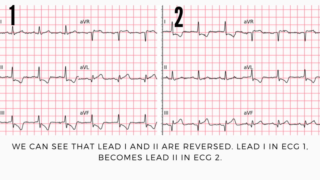
A few minutes later the ECG is repeated and looks quite different. What does the ECG below show and why does it look so different?

(1)
These 2 ECGs are quite different to each other. This second ECG shows, STE in III and aVF and STD and T wave inversion in I, aVL and STD in V2, and V3. This ECG shows a STEMI and this patient went to the Cath Lab.
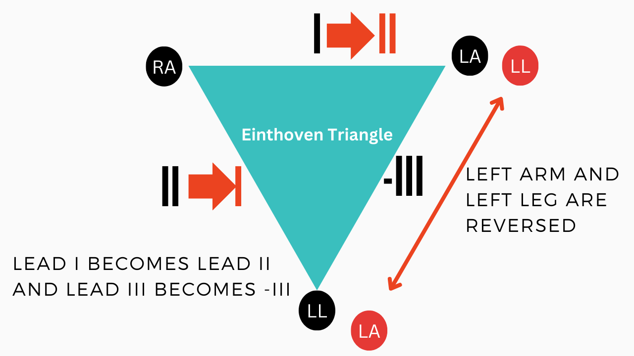
What has occurred here is lead misplacement of left arm and left leg.
Remember Einthoven’s triangle.

When the limb leads are reversed, lead I becomes lead II and lead III becomes -III. The precordial leads stay the same.
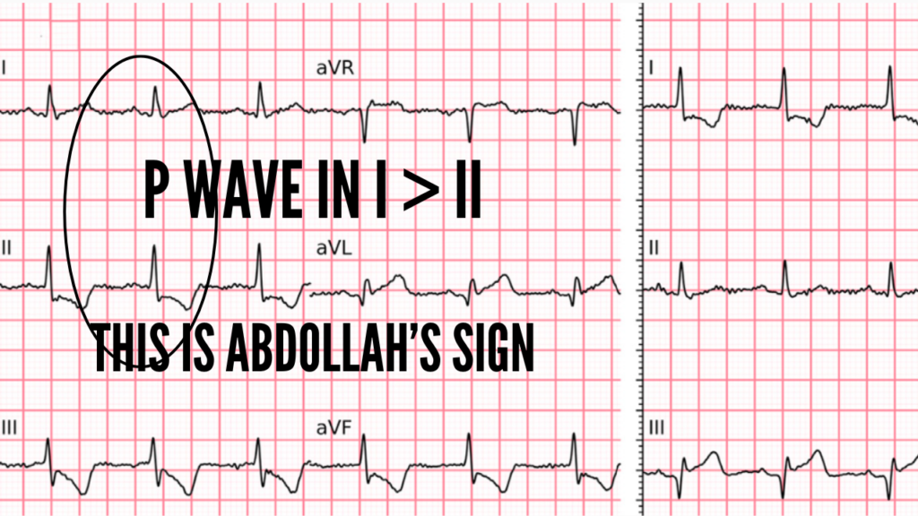
Abdollah Sign
Abdollah’s Sign is a non-specific, but potentially useful sign. We would normally expect the p wave to be taller in II ie II > I. Abdollah’s sign shows the opposite and alerts us to potential reversal of the arm and leg leads.
References
- Nunes de Alencar J. Annals of Emergency Medicine. November 2024: Vol 85. No 5pp 579-582


