A 19 yo male presents to the emergency department with palpitations. He has a narrow complex tachycardia on the monitor at a rate of 155 beats per minute. He has some left sided chest discomfort which this is pinpoint, sharp and totally reproducible. He states that he has had this for the last 2 days following working out at the gym.
An ECG is done and you diagnose SVT. You discuss what SVT means with the patient and progress to vagal manoeuvres, which don’t revert the patient. You then discuss the use of Adenosine with the patient. The patient reverts with a single dose of Adenosine.
The post reversion ECG is shown below. What does it show?
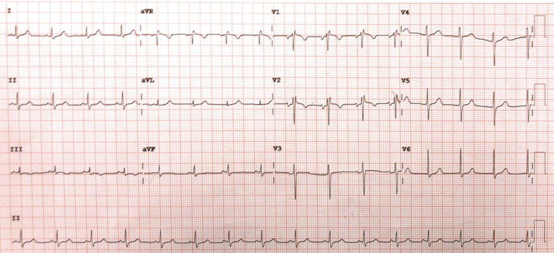
What is the Diagnosis?
(a) Anterior ST elevation Infarction
(b) Pulmonary Embolism
(c) Brugada Syndrome
(d) Lead misplacement
(e) Arrhythmogenic Right Ventricular Dysplasia
(a) Anterior ST elevation Infarction
(b) Pulmonary Embolism
(c) Brugada Syndrome
(d) Lead misplacement
(e) Arrhythmogenic Right Ventricular Dysplasia
ANSWER
(d) Lead misplacement
The ECG shows a RBBB pattern in V1 with inverted T waves in V1-2.
The p waves are the key here. The p wave in V1 is inverted and biphasic in V2. This indicates that these two leads are placed too high.
The ECG shows a RBBB pattern in V1 with inverted T waves in V1-2.
The p waves are the key here. The p wave in V1 is inverted and biphasic in V2. This indicates that these two leads are placed too high.