CASE
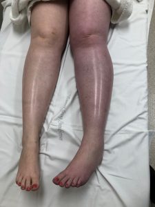 A 35 yo woman presents with a painful blue swollen leg, that occurred rapidly, the evening before. There is little relevant past history and the patient is on the oral contraceptive pill.
Examination reveals a dusky, blue left leg, that is swollen in comparison to the other side. Pulses are present and the patient in neurologically intact.
An ultrasound is performed and demonstrates an occlusive DVT that extends through the whole limb. It extends into the left common iliac, external iliac, common femoral and superficial femoral veins, as well as into the popliteal vein, extending into the calf.
The diagnosis is Phlegmasia Cerulean Dolens. Phlegmasia comes from the Greek Phlegma, which means inflammation, Cerulean from the Latin Ceruleus, meaning blue and Dolens from the Latin meaning painful.
The Hard Facts
This condition results from severe thrombotic occlusion of both major and collateral veins of an extremity. It predominantly affects the lower limbs and is more likely to affect the left limb, as in this case. The congestion can progress to compartment syndrome of the leg and is a precursor to gangrene, with significant amputation rates being reported.
Patients with this condition are also predisposed to massive pulmonary embolism, which occurs in up to 30% of cases, even when anti-coagulated.
The overall mortality approaches 40%.
Aetiology
There is an association with malignancy in about 40% of cases and patients will need to be investigated for this, especially for abdomen-pelvic lesions.
The condition is also associated with hyper-coagulable states and IVC filters.
Investigations
The investigation of choice is contrast venography, however duplex ultrasonography is the investigation most commonly used.
Other investigations that may be appropriate will include imaging to exclude malignancy or pathology testing to look at hypercoagulable states.
Management
Initially anticoagulation and a raising of the leg is needed. In some cases, if the cyanosis improves with elevation, simple anticoagulation may be considered. This can be LMWH, or oral factor Xa inhibitors. However due to the risk of serious complications with this condition, catheter-directed thrombolysis is the preferred treatment.
Our Case cont.
Following LMWH, our patient’s limb did improve in colour, marginally. The patient was transferred for catheter directed thrombolysis and further investigation.
Peter Kas