A 48 yo patient presents to the emergency department with a complaint of a sudden right temporal headache on exertion(exercising) 4 days previously that has now migrated to the right temporal region. This blog was first published on www.emmastery.com.
4 days previously whilst working out the patient had a sudden onset of severe frontal headache. The patient describes this as a 10 out of 10 headache that reached its peak within seconds.There was some associated mild photophobia and the patient was nauseated, but there was no emesis. Intitially the headache was bilateral, frontal and pulsatile. Over the next 4 days the headache has moved to the right temporal region and the patient complained of a small area over the right right temporal region that was painful to palpation. Mild photophobia persisted.
The patient had no past medical history of note. He was a non-smoker and drank socially. He did not take illicit substances.
On examination, the patient had normal vitals. He was afebrile, with a heart rate of 64 beats per minute and a blood pressure of 138/56 and oxygen saturations of 96% on room air.
He was oriented to person place and time, with normal speech and memory function. There was no neck stiffness. Cranial nerves were normal, as were visual fields. Upper and lower limb examination was essentially normal.
There was some tenderness to palpation over the right temporal region.
The patient had basic blood workup with a FBC, EUC, LFT, CRP and ESR, which was essentially normal.
 What is the definition of a thunderclap headache?
The International Headache Society describes it as: “headache that is severe in intensity, abrupt in onset (peaking in intensity in less than 1 minute), lasts at least 5 minutes, and is not accounted for by another diagnosis.
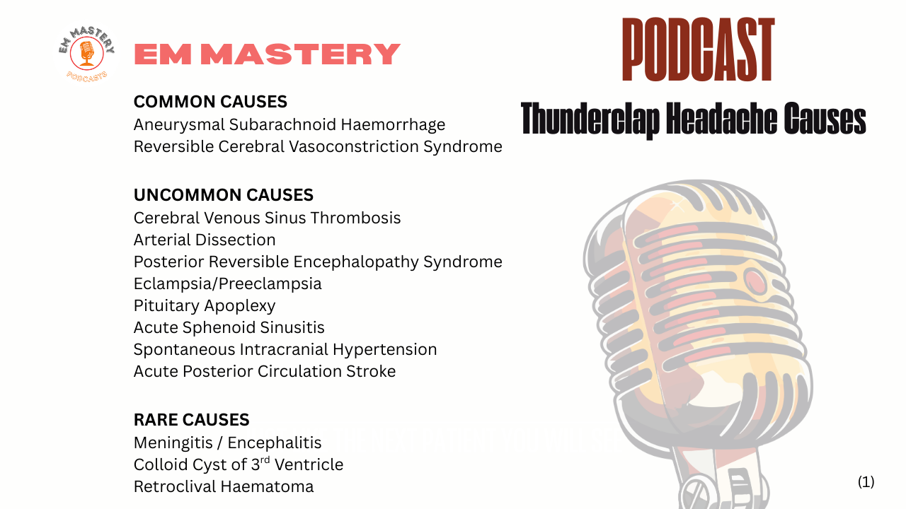
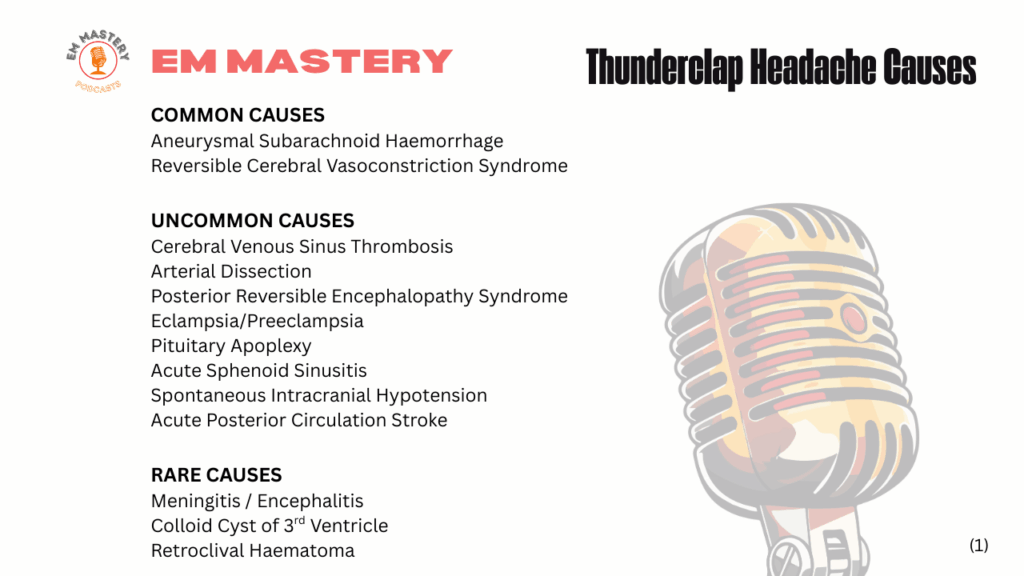
What are the Differential Diagnoses of a Thunderclap Headache?
1.Aneurysmal Subarachnoid Heamorrhage
Thunderclap headaches also known as warning leak headaches associated with aneurysmal subarachnoid haemorrhage peak within 1 minute of onset. True or False?
False: 15% of patients with an aneurysmal subarachnoid haemorrhage have headache onset peak after 1 minute and up to 60 minutes.
Can patients with aneurysmal subarachnoid haemorrhage present with only headache?
Up to 7% of patients present wityh a solitary thunderclap headache and no neurology.
Is a CT Non-contrast scan of the brain an adequate investigation to make a diagnosis of subarachnoid haemorrhage?
A CT performed within 6 hours of headache onset has a high sensitivity and can be used to rule out a subarachnoid haemorrhage.
We cannot rule out subarachnoid haemorrhage after 6 hours. The sensitivity of the scan falls significantly over time. An aneurysm needs to be ruled out by CT angiography.
Specifics to look for in the presentation
- Hypertension
- Neck pain / meningism
- Loss of Consciousness
- Vomiting
- Retinal Haemorrhage
- Look for CN III being affected
Investigations
- CT Brain if presenting < 6 hours after onset
- If presenting > 6 hours. Lumbar puncture or CT angiogram, which is becoming more common (read the review of current practice in suspected subarachnoid haemorrhage in The SHED Study)
- If Cavernous Venous Sinus Thrombosis (CVST) (see below is a differential ensure that the venous sinuses are looked at. This may require a dedicated CT venogram, however can be viewed on angiogram.
More Uncommon Causes of Thunderclap Headache
2.Reversible Cerebral Vasoconstriction Syndrome (RVCS)
It can present with a thunderclap headache and is associated with an emotional, physical or medical trigger. It is more common in women than men and patients usually report multiple thunderclap headaches over days or weeks.
The condition results in multifocal vasoconstriction of the intracranal arteries
Can RVCS cause a subarachnoid bleed?
About one third of patients with will have a convexal subarachnoid haemorrhage. It is characterised by reversible and fluctuating narrowing of the intracranial arteries.
Specifics to look for in the presentation
- There is a vasoconstrictive trigger(emotional, physical and even medical) prior to headaches
- Multiple thunderclap headaches over days to weeks is pathognomonic- However we need to remember that this may be the first headache
Investigations
-
CT angiography or MR angiography
- Beware as these may be normal early in the presentation.
3.Cerebral Venous Sinus Thrombosis (CVST)
Up to 15% of patients with CVST will present with a thunderclap headache.
Specifics to look for in the presentation
- It is more common in young adults (<50) and females
- The presentation depends on the location of the thrombus
- Risk factors include:
- Prothrombotic state
- Oestrogen intake
- Pregnancy especially 3rd trimester
- Head and Neck infections
- Thrombophilia
- Trauma, ie mechanical or iatrogenic(neurosurgical procedures)
- Prothrombotic state
- Patients can present with:
Visual problems including decreased vision and proptosis - Sensory and motor deficits
- Seizures
- Aphasia(Left Transverse sinus thrombosis)
Investigations
-
CT or MR venography
- Beware as these may be normal early in the presentation.
Listen to the 3 minute podcast on Cavernous Sinus Thrombosis given by Dr Joe Nemeth.
4.Arterial Dissection
Thunderclap headache has been reported in both carotid and vertebral artery dissections. It can also cause convexal subarachnoid hemorrhage.
(von Babo M, De Marchis GM, Sarikaya H, et al. Differences and similarities between spontaneous dissections of the
internal carotid artery and the vertebral artery. Stroke 2013; 44: 1537-42.)
Specifics to look for in the presentation
- Neck pain due to cervical artery dissection
- Background of trauma
- Headache may precede or occur with stroke symptoms.
- Look for a Horner’s Sydrome
Investigations
-
CT or MR angiography
5.Posterior Reversible Encephalopathy Syndrome
This can also cause severe headache, however the onset is not sudden. Patients also commonly have altered vision, seizures or an altered level of consciousness.
Specifics to look for in the presentation
- History of Hypertension
- Clinical presentation includes
- Visual loss
- Altered level of consiousness
- Seizures
Investigations
-
MRI
6.Pituitary Apoplexy
In Pituitary Apoplexy there is an acure infarct , or haemorrhage of the pituitary gland usually on the background of a pre-existing pituitary tumour. Precipitating factors can include pregnancy and anticoagulation.
Specifics to look for in the presentation
- Sudden headache usually behind the eyes.
- Clinical presentation includes
- Visual symptoms
- Decreased visual acuity
- Visual field defects
- Diplopia,
- Ptosis,
- Later eye deviation and mydriasis.
- Altered level of consiousness
- Seizures
- Visual symptoms
Investigations
-
MRI
- CT scan as the initial scan may be eaiser to obtain
7.Eclampsia and Pre-eclampsia
It is an uncommon cause of sudden severe headache, but patients may present with headaches. They may also present with scotomas and cortical blindness.
Specifics to look for in the presentation
- Pregnancy or first 12 weeks postpartum.
- Clinical presentation includes
- SBP > 140 mm Hg or DBP > 90 mm Hg
- Visual disturbance
- Seizures
- Proteinuria
- Hyper-reflexia
- Abdominal Pain
Investigations
-
Labs and Urine
-
MRI
8.Spontaneous Intracranial Hypotension
Patients can present with a sudden or gradual headache. It can be generalised or focal. It is usually postural and is relieved by lying down. It can be exacerbated by coughing or head movement. It is caused by a CSF leak that is eather congenital or traumatic and results in a tear in the dura. This leak results in the brain ‘sagging’ in the cranial vault and causing teraction on sensory nerve fibres.
Specifics to look for in the presentation
- Clinical presentation
- Postural headache is most common
- Diplopia and blurred vision
- Photophobia
- Neck stiffness
- Unsteady gait
Investigations
- MRI
- LP recording opening pressures
9.Giant Cell Arteritis
Patients with Giant Cell Artertiis(GCS) or Temporal Arteritis, are usually older (>50yo) present, in most cases, with temporal headache, although the headache can be in other areas or generalised. Symptoms may be acute in onset and last several days to weeks.
Specifics to look for in the presentation
- Headache
- Myalgia, fatigue and malaise
- Night sweats
- Jaw claudication
- Unexplained fever
- Pain over the scalp and/or over the temporal artery region.
- Eye signs
- Visual loss/blurred vision
- Visual field defects
- Diplopia
- Ptosis
Investigations
- Labs
- Raised ESR (usually > 50 mm/h- but can be normal in up to 20% of cases.
- CRP rises before ESR and has greater sensitivity for GCA
- Using both ESR and CRP provides a greater sensitivity.
- A full Blood count will show an elevated platelet count in most patients.
- Temporal Artery Biopsy
- Color Duplex Ultrasound, may be an alternative moving forward.
10.Primary Exertional Headache
Exercise-induced headaches may vary from mild to severe headaches, triggered by intense physical exertion and usually associted with resistance training, cycling or running, but can be associated with other types of sports.
The headaches are usually diffuse, but can be localised. They are usually throbbing in nature and tend to last for less than 48 hours.
The mean age of presentaion is 24 years. Older patients presenting with exercise-induced headaches, should raise suspicion for secondary causes of headache. These may include:
- Intracranial structural abnormalities
- Intracranial haemorrhage
- Neoplasm
- Space occupying Lesions such as cysts
- Chiari Malformations (Congenital anatomic abnormalities of the craniocervical junction)
- Intracranial Hypertension
- Vascular Causes
- Reversible Cerebral vasoconstriction Syndrome
- Carotid/Vertebral(Cervical) artery dissection
- Cardiac aetiology
- Ischaemia
- Hypertension
- Takotsubo
Specifics to look for in the presentation
Diagnostic criteria (ICHD-3) requires the following criteria to be presents to make the diagnosis:
- At least 2 headache episodes
- Occur only during strenuous exercise
- Last for <48 hours
- No other ICHD-3 diagnosis would better explain the headache.
Investigations
Sudden onset severe headache during exertion, should first rule out subarachnoid haemorrhage and cerebral aneurysm.
- CT brain and CTA (head and neck) or MRA .
- labs for other conditions including giant cell arteritis and cardiac ischaemia.
Plus listen to Sudden Headache:3 Diagnoses not to miss. on EM Mastery Podcasts
References
- Edlow J et al. Case 18-2024: A 64 – year old Woman with the Worst Headache of Her Life. NEJM 2024;390:2108-18.
- Headache Classification Committee of the International Headache Society (IHS). The International Classification of Headache Disorders, 3rd edition. Cephalalgia 2018; 38: 1-211.
- Perry JJ, et al. Clinical decision rules to rule out subarachnoid hemorrhage for acute headache. JAMA 2013; 310: 1248-55.
- Linn FH, et al. Headache characteristics in subarachnoid haemorrhage and benign thunderclap headache. J Neurol Neurosurg Psychiatry 1998; 65: 791-3.
- Roberts T, et al. Thunderclap headache syndrome presenting to the emergency department: an international multicentre observational cohort study. Emerg Med J 2022; 39: 803-9.
- Perry JJ, et al. Clinical decision rules to rule out subarachnoid hemorrhage for acute headache. JAMA 2013; 310: 1248-55.
- Perry JJ, et al. High risk clinical characteristics for subarachnoid haemorrhage in patients with acute headache: prospective cohort study. BMJ 2010; 341: c5204.
- Dubosh NM, et al. Sensitivity of early brain computed tomography to exclude aneurysmal subarachnoid hemorrhage: a systematic review and meta-analysis. Stroke 2016; 47: 750-5.
- Ducros A. Reversible cerebral vasoconstriction syndrome. Lancet Neurol 2012;11: 906-17.
- Singhal AB, et al. Reversible cerebral vasoconstriction syndromes: analysis of 139 cases. Arch Neurol 2011; 68: 1005-12.
- Topcuoglu MA, et al. Hemorrhagic reversible cerebral vasoconstriction syndrome: features and mechanisms. Stroke 2016; 47: 1742-7.
- Cumurciuc R, et al. Headache as the only neurological sign of cerebral venous thrombosis: a series of 17 cases. J Neurol Neurosurg Psychiatry 2005; 76: 1084-7.
- de Bruijn SF, et al. Thunderclap headache as first symptom of cerebral venous sinus thrombosis. Lancet 1996; 348: 1623-5.
- Refai D, et al. Spontaneous isolated convexity subarachnoid hemorrhage: presentation, radiological findings, differential diagnosis, and clinical course. J Neurosurg 2008; 109:1034-41.
- Pascual J, et al. Cough, exertional, and sexual headaches: an analysis of 72 benign and symptomatic cases. Neurology 1996; 46:1520.
- Headache Classification Committee of the International Headache Society (IHS) The International Classification of Headache Disorders, 3rd edition. Cephalalgia 2018; 38:1.